-
Non Invasive Imaging Directly On Your Microscope
Quantitative, real time, 3D imaging of cell, tissue, and organoids assays, un-cleared small model animals and plants -
Non Invasive Imaging Directly On Your Microscope
Quantitative, real time, 3D imaging of cell, tissue, and organoids assays, un-cleared small model animals and plants
![]()
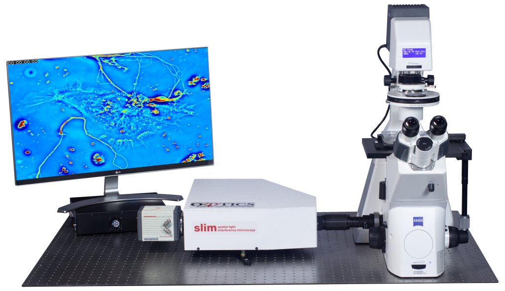
Cell assays, protein particles, and
digital pathology slides
![]()
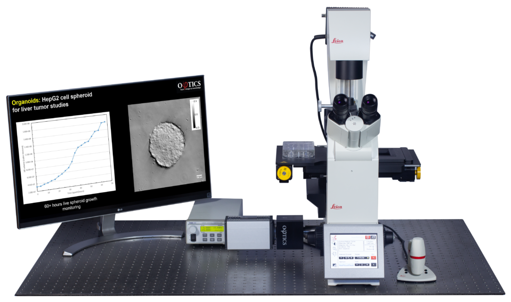
Organoid and tissue assays,
embryos, cell clusters
![]()
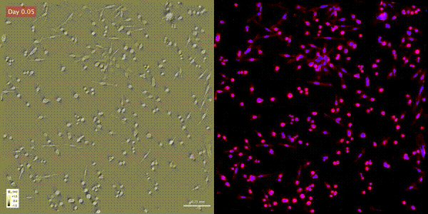
Live digital staining of CellVista SLIM
and CellVista GLIM assays
Benefits of QPI for Live Cell Microscopy
Quantitative Phase Imaging (QPI) is a new imaging channel that measures the intrinsic properties (dry mass, refractive index) and topography of biological specimens. Phi Optics’ QPI is the fastest and most accurate imaging of live cell and tissue cultures, providing contrast and resolution where regular microscopy fails or kills the specimens. Phi Optics QPI upgrades your existing microscopes to enable 4D non-destructive, quantitative assays on any range of time, with subcellular resolution.
Scientific Articles
2026
2024
News & Events
Quantitative-mass imaging measure fluctuations during bacterial growth with greater precision
Researchers used CellVista SLIM and new advanced microfluidics to reveal cell-to-cell variability in dry density and density fluctuations during bacterial growth. With greater precision and throughput, the results suggest a potential homeostasis mechanism and related effects on bacterial replication.
See the science: https://www.nature.com/articles/s42003-022-03348-2
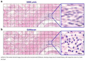
Continuous scanning microscopy uses deep learning deblurring
Want high imaging throughput without motion blurring? Use deep learning deblurring #GANScan and recover the original image resolution. To read more about the science, please visit https://www.nature.com/articles/s41377-022-00952-z
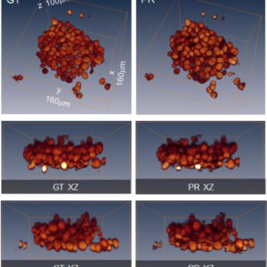
AI meets label-free cell imaging through artificial confocal microscopy
Scientists at University of Illinois at Urbana Champaign and Washington University in St. Louis combined CellVista GLIM® with confocal illumination and deep learning PICS to segment individual nuclei within dense spheroids for both cell counting and volume measurements without tags!
To read more about the science, please visit https://www.nature.com/articles/s41566-022-01140-6.
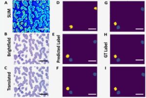
Breakthrough in blood smear evaluations can lead to faster test results
Scientists at University of Illinois at Urbana Champaign, Christie Clinic and Washington University in St. Louis used #cSLIM coupled with #PICS to classify white blood cells with 75% mean average precision – opening the door for rapid and automated QPI evaluations of unlabeled clinical blood smears.
To read more about the science, please visit: https://www.nature.com/articles/s41598-022-21250-z