Automatic Colorectal Cancer Screening Using Deep Learning in Spatial Light Interference Microscopy Data
J.K. Zhang, M. Fanous, N. Sobh, A. Kajdacsy-Balla, and G. Popescu
Cells 2022 11(4) 716 https://doi.org/10.3390/cells11040716
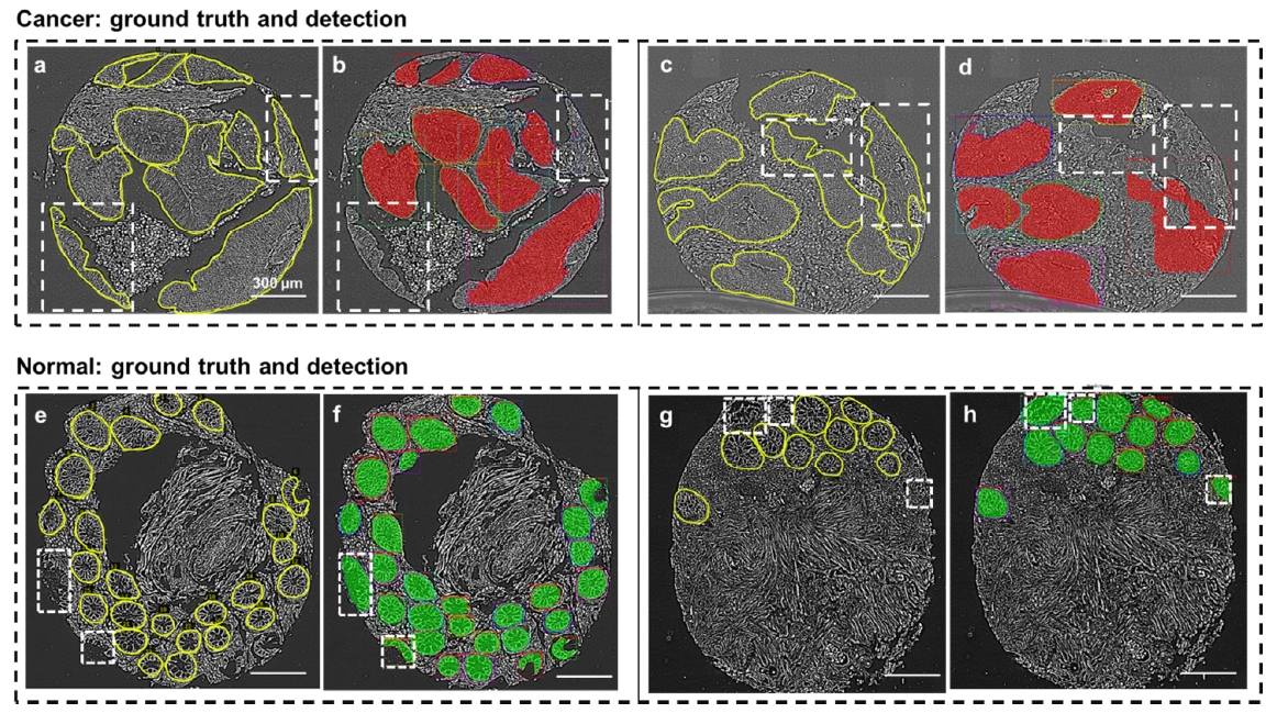
The surgical pathology workflow currently adopted by clinics uses staining to reveal tissue architecture within thin sections. A trained pathologist then conducts a visual examination of these slices and, since the investigation is based on an empirical assessment, a certain amount of subjectivity is unavoidable. Furthermore, the reliance on external contrast agents such as hematoxylin and eosin (H&E), albeit being well-established methods, makes it difficult to standardize color balance, staining strength, and imaging conditions, hindering automated computational analysis. In response to these challenges, we applied spatial light interference microscopy (SLIM), a label-free method that generates contrast based on intrinsic tissue refractive index signatures. Thus, we reduce human bias and make imaging data comparable across instruments and clinics. We applied a mask R-CNN deep learning algorithm to the SLIM data to achieve an automated colorectal cancer screening procedure, i.e., classifying normal vs. cancerous specimens. Our results, obtained on a tissue microarray consisting of specimens from 132 patients, resulted in 91% accuracy for gland detection, 99.71% accuracy in gland-level classification, and 97% accuracy in core-level classification. A SLIM tissue scanner accompanied by an application-specific deep learning algorithm may become a valuable clinical tool, enabling faster and more accurate assessments by pathologists.