White Blood Cell Detection, Classification and Analysis Using Phase Imaging with Computational Specificity (PICS)
Michael J Fanous, Shenghua He, Sourya Sengupta, Krishnarao Tangella, Nahil Sobh, Mark A Anastasio, Gabriel Popescu
Scientific Reports 2022 12 20043 https://doi.org/10.1038/s41598-022-21250-z
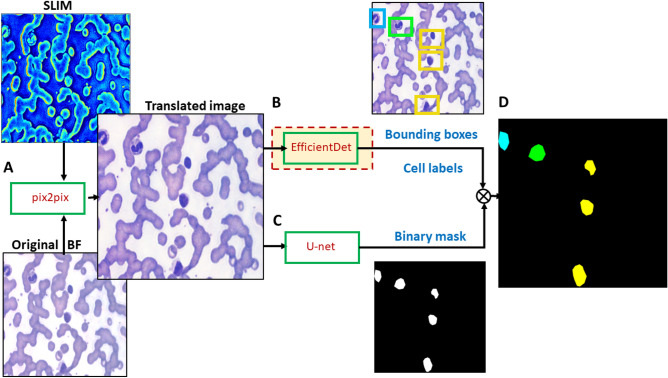
Treatment of blood smears with Wright’s stain is one of the most helpful tools in detecting white blood cell abnormalities. However, to diagnose leukocyte disorders, a clinical pathologist must perform a tedious, manual process of locating and identifying individual cells. Furthermore, the staining procedure requires considerable preparation time and clinical infrastructure, which is incompatible with point-of-care diagnosis. Thus, rapid and automated evaluations of unlabeled blood smears are highly desirable. In this study, we used color spatial light interference microcopy (cSLIM), a highly sensitive quantitative phase imaging (QPI) technique, coupled with deep learning tools, to localize, classify and segment white blood cells (WBCs) in blood smears. The concept of combining QPI label-free data with AI for the purpose of extracting cellular specificity has recently been introduced in the context of fluorescence imaging as phase imaging with computational specificity (PICS). We employed AI models to first translate SLIM images into brightfield micrographs, then ran parallel tasks of locating and labelling cells using EfficientNet, which is an object detection model. Next, WBC binary masks were created using U-net, a convolutional neural network that performs precise segmentation. After training on digitally stained brightfield images of blood smears with WBCs, we achieved a mean average precision of 75% for localizing and classifying neutrophils, eosinophils, lymphocytes, and monocytes, and an average pixel-wise majority-voting F1 score of 80% for determining the cell class from semantic segmentation maps. Therefore, PICS renders and analyzes synthetically stained blood smears rapidly, at a reduced cost of sample preparation, providing quantitative clinical information.