Digital staining facilitates biomedical microscopy
Michael John Fanous, Nir Pillar, and Aydogan Ozcan
Front Bioinform. 2023 3 1243663 https://doi.org/10.3389/fbinf.2023.1243663
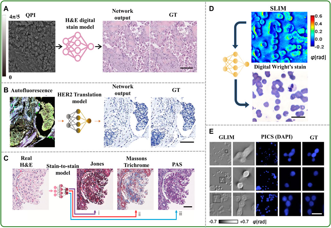
Traditional staining of biological specimens for microscopic imaging entails time-consuming, laborious, and costly procedures, in addition to producing inconsistent labeling and causing irreversible sample damage. In recent years, computational “virtual” staining using deep learning techniques has evolved into a robust and comprehensive application for streamlining the staining process without typical histochemical staining-related drawbacks. Such virtual staining techniques can also be combined with neural networks designed to correct various microscopy aberrations, such as out-of-focus or motion blur artifacts, and improve upon diffracted-limited resolution. Here, we highlight how such methods lead to a host of new opportunities that can significantly improve both sample preparation and imaging in biomedical microscopy.