Spatial Light Interference Microscopy — High Contrast Imaging for Optically Thin Specimens
CellVista-SLIM features:
CellVista-SLIM upgrades your optical microscopes for 4D label-free quantitative imaging:
- Quantitative cell assays: growth and proliferation, cytotoxicity, viability, immune cell killing, senescence
- Digital pathology slides: tissue morphology and refractive index mapping with or without staining
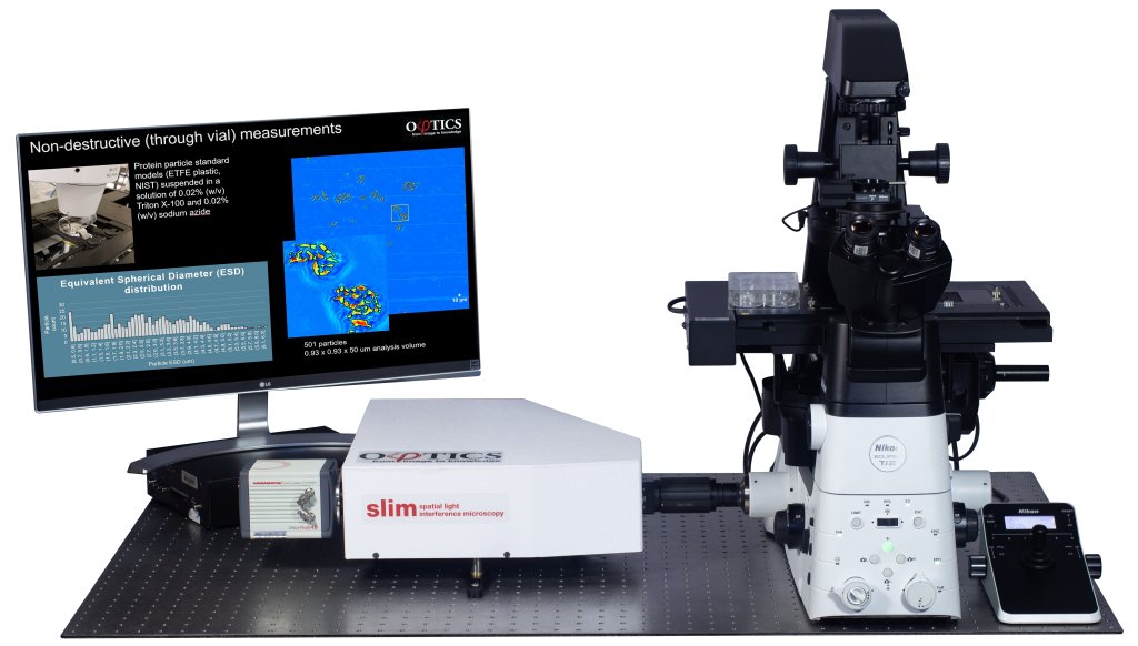
- Size, count and 3D morphology of protein particles
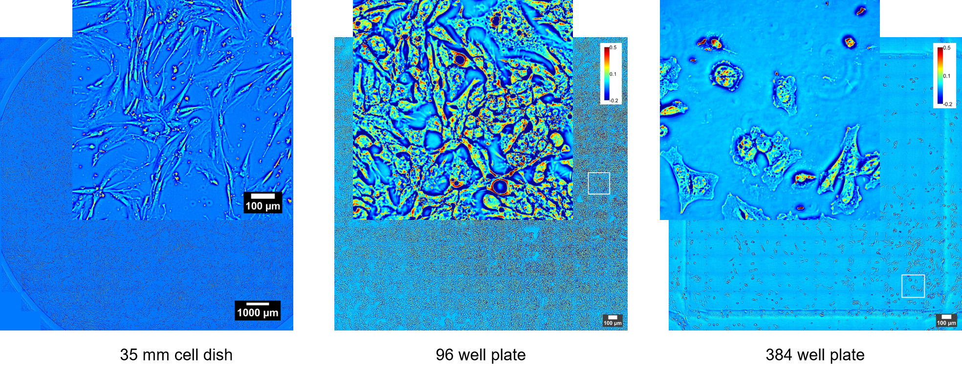
CellVista SLIM upgrades the camera port of Phase Contrast commercial microscopes (Zeiss, Nikon, Leica, Olympus) with any magnification available (immersion or dry, 10x to 100x). Samples can be loaded in standard holders (glass slides, single and multi-well plates) and fields of view are limited only by the microscope stage movement. High end sCMOS cameras are integrated for high sensitivity fluorescence co-localization.
CellVista SLIM
Advantages
Advantages
+ High throughput: image large cell populations with single cell resolution for multiple ranges of time
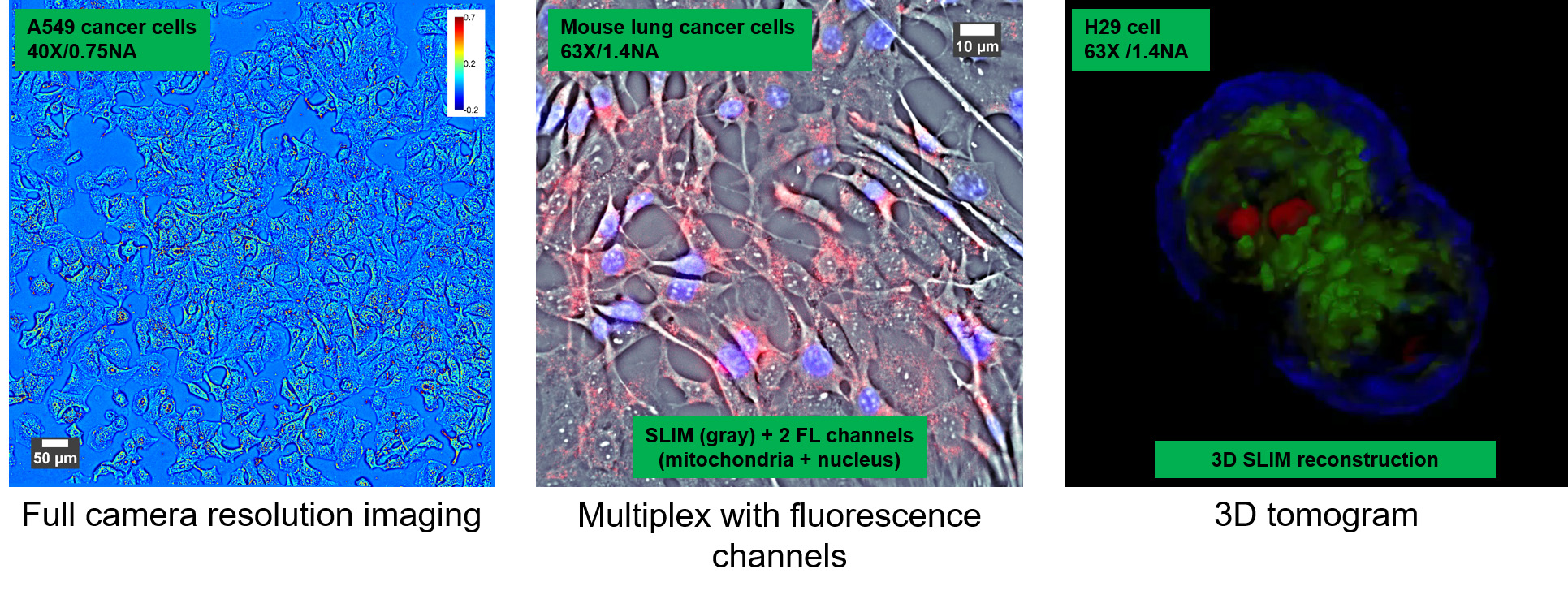
+ High sensitivity: no speckles, low background to detect sub-cellular features
+ Correlative imaging with any fluorescence channels: quantify with SLIM and identify with fluorescence!
+ 3D tomography of entire field of view
+ High sensitivity: no speckles, low background to detect sub-cellular features
+ Correlative imaging with any fluorescence channels: quantify with SLIM and identify with fluorescence!
+ 3D tomography of entire field of view
For a 3D tomography solution for thick specimens please see our CellVista GLIM™ systems. For customization don’t hesitate to CONTACT US!