Gradient Light Interference Microscopy — High Contrast Imaging for Optically Thick Specimens

CellVista-GLIM features:
CellVista-GLIM upgrades your optical microscopes for 4D label-free quantitative imaging:
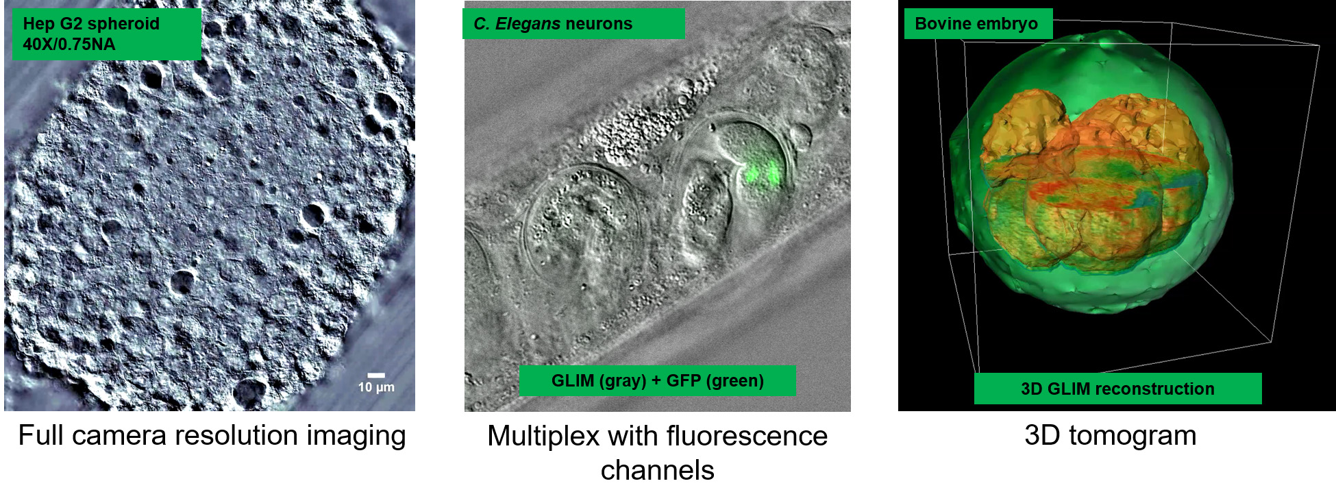
- 3D morphology of embryos and cell clusters
- 3D organoid assays with single cell resolution
- Animal and plant histology
- Microscopic animal models
CellVista GLIM upgrades the camera port of DIC commercial microscopes (Zeiss, Nikon, Leica, Olympus) with any magnification available (immersion or dry, 10X to 100X). Samples can be loaded in standard holders (DIC-compatible glass slides, glass-bottom single and multi-well plates) and fields of view are limited only by the microscope stage movement. High end sCMOS cameras are integrated for high sensitivity fluorescence co-localization.
CellVista GLIM
Advantages
Advantages
+ High throughput: image 3D specimens with single cell resolution for multiple ranges of time
+ High sensitivity: reduce scattering background for 3D tomography of 100s microns thick specimens!
+ Correlative imaging with any fluorescence channels: quantify with GLIM and identify with fluorescence!
+ High sensitivity: reduce scattering background for 3D tomography of 100s microns thick specimens!
+ Correlative imaging with any fluorescence channels: quantify with GLIM and identify with fluorescence!
For high image resolution solutions of thin specimens please see our CellVista SLIM™ systems. For customization don’t hesitate to CONTACT US!