ZINC’S EFFECT ON THE DIFFERENTIATION OF PORCINE ADIPOSE-DERIVED STEM CELLS INTO OSTEOBLASTS
Josh Bertels1 , Marcello Rubessa1 , Mikhail E Kandel2 , Thomas Bane1 , Derek J Milner1 , Gabriel Popescu2,3, and Matthew B Wheeler1,3,4*
Journal of Regenerative Medicine, 8, 2 2019
![]()

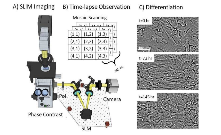
The aim of this project was to evaluate the effects of zinc in osteogenic media on differentiation of adipose-derived stem cells (ASC) into osteoblasts. ASC were plated (50,000 cells/mL) and subjected to treatments of zinc at six different concentrations in osteogenic medium: 8 mM, 4 mM, 0.8 mM, 0.4 mM, 0.08 mM, and 0.04 mM, as well as standard osteogenic medium as a positive control, and ASC culture medium as a negative control. At the end of the 4-week incubation period, cultures were analyzed for osteogenic differentiation, as measured by Alizarin Red S staining, osteocalcin production and Spatial Light Interference Microscopy (SLIM). Here we demonstrate that zinc supplementation to a standard ASC osteogenesis-promoting media formulation has a positive effect on the differentiation of porcine ASC into osteoblasts, and that 0.08 mM is the optimum concentration. Average size and number of osteogenic nodules increased under zinc supplementation, cells changed morphology more rapidly, and the concentration of osteocalcin released during osteogenic differentiation was higher (p=0.0004). Additionally we have demonstrated that a new noninvasive method utilizing SLIM microscopy can provide researchers another tool to probe intracellular and intracultural behavior in vitro, linking changes in cell behavior to differences in culture media formation. This specialized microscopic visualization technique may provide researchers with a good, non-invasive tool to evaluate the effect of molecular additives to media formulations on cell behavior, including potentially screening the effect of small molecule compounds derived from chemical libraries on cellular behavior in vitro.

