Quantitative Phase Imaging of Stromal Prognostic Markers in Pancreatic Ductal Adenocarcinoma
Fanous M, Keikhosravi A, Kajdacsy-Balla A, Eliceiri KW, Popescu G.
Biomedic Optics Express 2020 11(3) 1354 https://doi.org/10.1364/BOE.383242
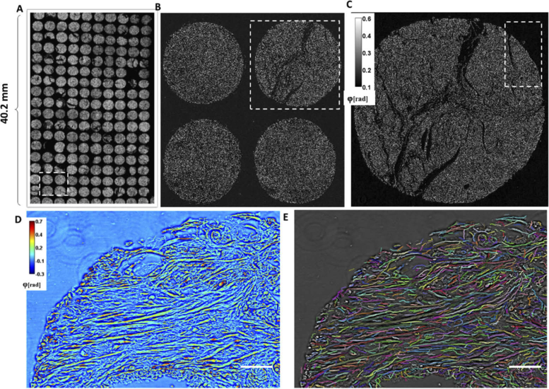
New quantitative prognostic markers are needed for improved pancreatic ductal adenocarcinoma (PDAC) prognosis. Second harmonic generation microscopy has been used to show that collagen fiber alignment in PDAC is a negative prognostic factor. In this work, a series of PDAC and normal adjacent tissue (NAT) biopsies were imaged with spatial light interference microscopy (SLIM). Quantitative analysis performed on the biopsy SLIM images show that PDAC fiber structures have lower alignment per unit length, narrower width, and are longer than NAT controls. Importantly, fibrillar collagen in PDAC shows an inverse relationship between survival data and fiber width and length (p < 0.05).