QUANTITATIVE PHASE IMAGING IN BIOMEDICINE
YongKeun Park1,2,3, Christian Depeursinge4,5 and Gabriel Popescu 6*
Nature Photonics, VOL 12, Pages 578–589 2018 https://doi.org/
![]()
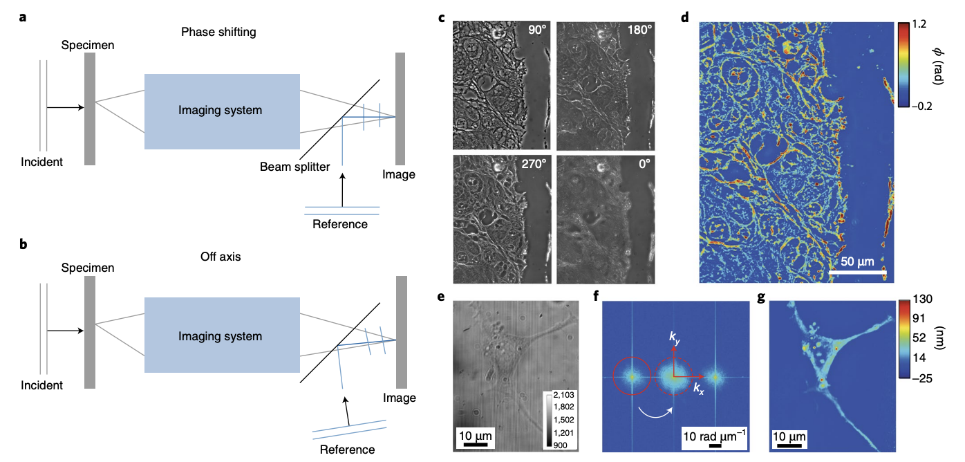
Quantitative phase imaging (QPI) has emerged as a valuable method for investigating cells and tissues. QPI operates on unlabelled specimens and, as such, is complementary to established fluorescence microscopy, exhibiting lower phototoxicity and no photobleaching. As the images represent quantitative maps of optical path length delays introduced by the specimen, QPI provides an objective measure of morphology and dynamics, free of variability due to contrast agents. Owing to the tremendous progress witnessed especially in the past 10–15 years, a number of technologies have become sufficiently reliable and translated to biomedical laboratories. Commercialization efforts are under way and, as a result, the QPI field is now transitioning from a technology-development-driven to an application-focused field. In this Review, we aim to provide a critical and objective overview of this dynamic research field by presenting the scientific context, main principles of operation and current biomedical applications.