Label-free correlative morpho-chemical tomography of 3D kidney mesangial cells
Butola A, Ghosh B, Park J, Kwon M, De la Cadena A, Mukherjee SS, Bhargava R, Boppart SA, and Agarwal K.
J Biomed Opt. 2026 31(3) 036501 https://doi.org/10.1117/1.JBO.31.3.036501
![]()
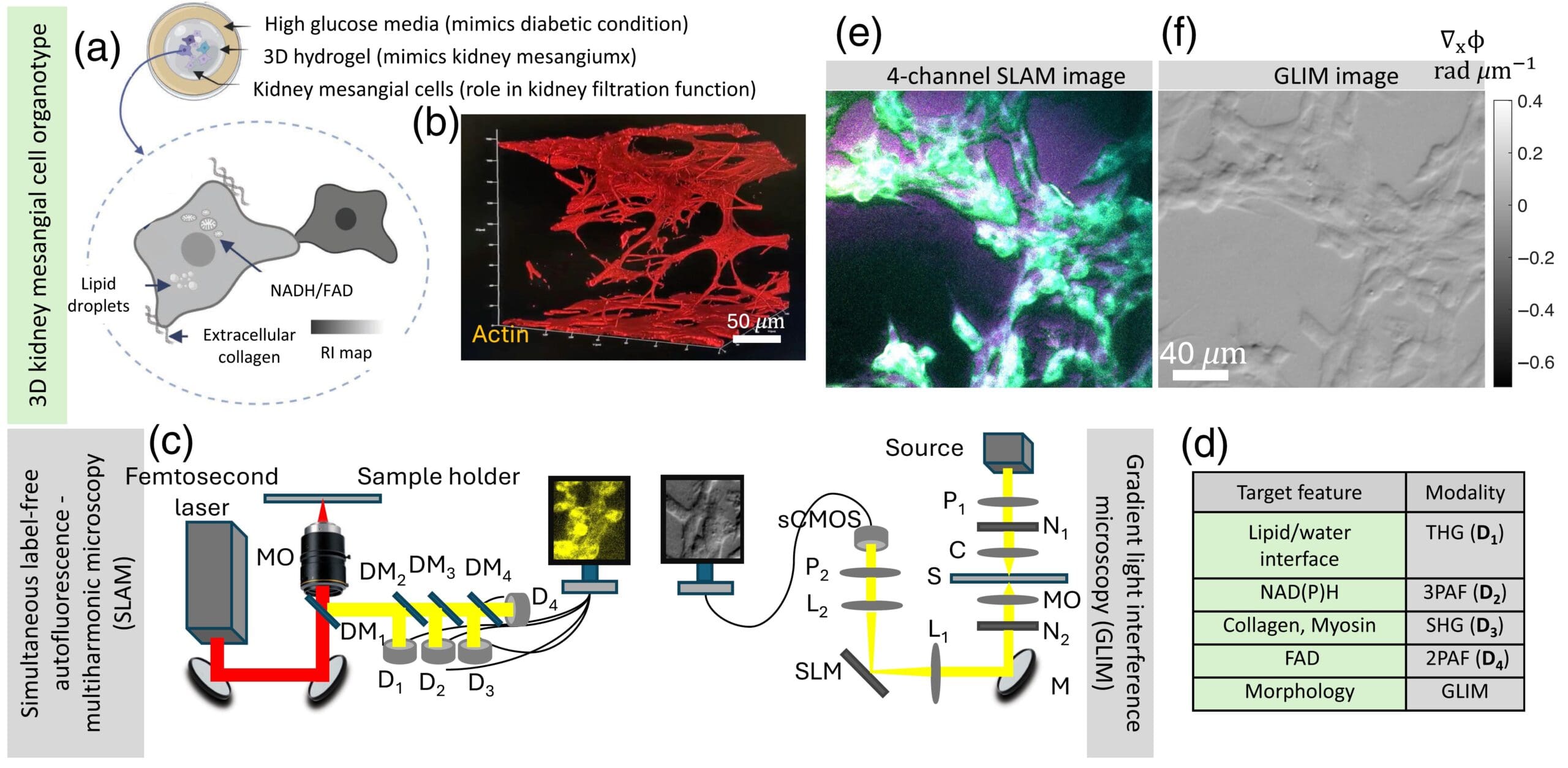
Significance: Imaging 3D in vitro kidney models is essential to understand kidney function and pathology. Label-free characterization of such specimens seeks to supplement existing imaging techniques and avoid the need for contrast agents that can disturb the native state of living samples. Conventional label-free optical imaging techniques are compatible with living samples but face challenges such as poor sectioning capability, fragmentary morphology, and lack of chemical-specific information. Aim: We aim to develop and demonstrate a correlative label-free imaging platform capable of simultaneously capturing morphological and chemical-specific information from 3D cultured kidney mesangial cells. Approach: We combined simultaneous label-free autofluorescence-multiharmonic (SLAM) microscopy and gradient light interference microscopy (GLIM) to extract both chemical-specific and morphological tomography of 3D cultured kidney mesangial cells. In this approach, SLAM provides a nonlinear imaging platform with a single excitation source to simultaneously acquire autofluorescence (FAD and NAD(P)H), second- and third-harmonic signals from the cells. Complementarily, GLIM acquires high-contrast quantitative phase information to quantify structural changes in samples with a thickness of up to 250 μm. Results: Our correlative imaging results demonstrate the ability to image and quantify both morphology and chemical-specific signals of kidney mesangial cells in 3D. The combination of GLIM and SLAM provides complementary information critical for understanding kidney function, including metabolism and matrix deposition under controlled physiological conditions. Conclusions: The proposed correlative imaging approach establishes a versatile and hassle-free platform for morpho-chemical cellular tomography, offering unique opportunities for studying the structure and function of 3D kidney models in their native state.