IMAGING COLLAGEN PROPERTIES IN THE UTEROSACRAL LIGAMENTS OF WOMEN WITH PELVIC ORGAN PROLAPSE USING SPATIAL LIGHT INTERFERENCE MICROSCOPY (SLIM)
Chenfei Hu1,2,3, Melissa Santi 3 , Oluwatobi Adelaja4 , Andre Kajdacsy-Balla4 , Gabriel Popescu1,2,3 and William Kobak 5 *
Optics and Photonics, Frontiers in Physics, 10, Page 3389 2019
![]()
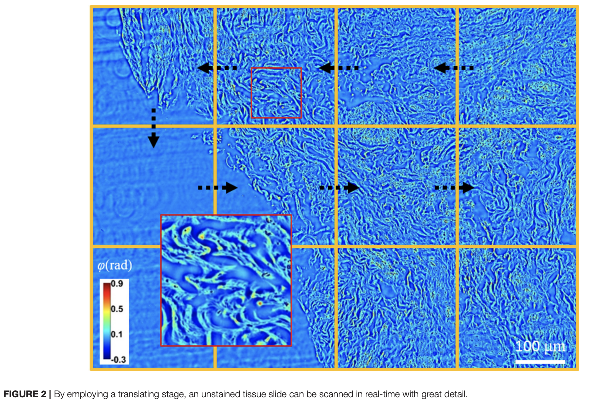
We studied collagen fiber organization in tissue affected by pelvic organ prolapse (POP) and compared it to asymptomatic controls. Both the control and POP tissue biopsies were prepared and measured by a highly sensitive quantitative phase imaging (QPI) system, called spatial light interference microscopy (SLIM). Combined with automatic image processing, this modality provides quantitative, high-throughput assessment of fiber morphology. We found the fiber orientation in prolapsed specimens is less homogeneous, indicating an abnormal organization of collagen in the extracellular matrix (ECM).