FOURIER PHASE MICROSCOPY WITH WHITE LIGHT, BIOMED. OPT. EXP. 4 (8), 1434-1441 (2013).

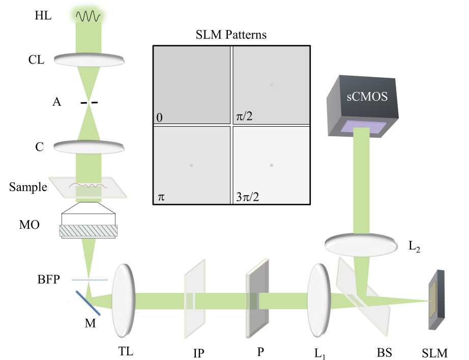
Laser-based Fourier phase microscopy (FPM) works on the principle of decomposition of an image field in two spatial components that can be controllably shifted in phase with respect to each other. However, due to the coherent illumination, the contrast in phase images is degraded by speckles. In this paper we present FPM with spatially coherent white light (wFPM), which offers high spatial phase sensitivity due to the low temporal coherence and high temporal phase stability due to common path geometry. Further, by using a fast spatial light modulator (SLM) and a fast scientific-grade complementary metal oxide semiconductor (sCMOS) camera, we report imaging at a maximum rate of 12.5 quantitative phase frames per second with 5.5 mega pixels image size. We illustrate the utility of wFPM as a contrast enhancement as well as dynamic phase measurement method by imaging section of benign colonic glands and red blood cell membrane fluctuation.
