EPI-Illumination Gradient Light Interference Microscopy for Imaging Opaque Structures
Mikhail E. Kandel, Chenfei Hu, Ghazal Naseri Kouzehgarani, Eunjung Min, Kathryn Michele Sullivan, Hyunjoon Kong, Jennifer M. Li, Drew N. Robson, Martha U. Gillette, Catherine Best-Popescu & Gabriel Popescu
Nat. Communications 2019 10 4691 https://doi.org/10.1038/s41467-019-12634-3
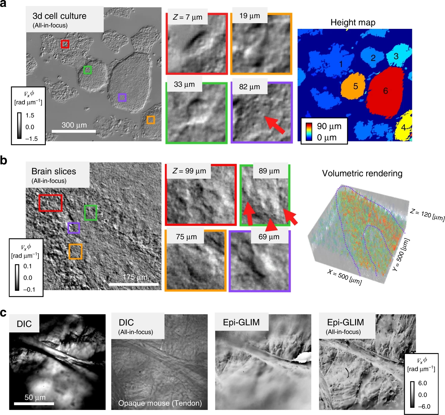
Multiple scattering and absorption limit the depth at which biological tissues can be imaged with light. In thick unlabeled specimens, multiple scattering randomizes the phase of the field and absorption attenuates light that travels long optical paths. These obstacles limit the performance of transmission imaging. To mitigate these challenges, we developed an epi-illumination gradient light interference microscope (epi-GLIM) as a label-free phase imaging modality applicable to bulk or opaque samples. Epi-GLIM enables studying turbid structures that are hundreds of microns thick and otherwise opaque to transmitted light. We demonstrate this approach with a variety of man-made and biological samples that are incompatible with imaging in a transmission geometry: semiconductors wafers, specimens on opaque and birefringent substrates, cells in microplates, and bulk tissues. We demonstrate that the epi-GLIM data can be used to solve the inverse scattering problem and reconstruct the tomography of single cells and model organisms.