DIFFRACTION PHASE AND FLUORESCENCE MICROSCOPY, OPT. EXP. 14, 8263, (2006)
Y. K. PARK, G. POPESCU, K. BADIZADEGAN, R. R. DASARI AND M. S. FELD
2006
![]()

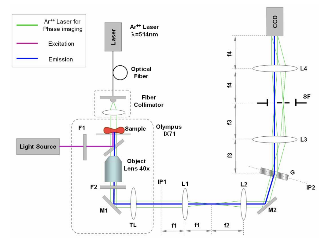
We have developed diffraction phase and fluorescence (DPF) microscopy as a new technique for simultaneous quantitative phase imaging and epi-fluorescence investigation of live cells. The DPF instrument consists of an interference microscope, which is incorporated into a conventional inverted fluorescence microscope. The quantitative phase images are characterized by sub-nanometer optical path-length stability over periods from milliseconds to a cell lifetime. The potential of the technique for quantifying rapid nanoscale motions in live cells is demonstrated by experiments on red blood cells, while the composite phase-fluorescence imaging mode is exemplified with mitotic kidney cells.
