Computational Interference Microscopy Enabled by Deep Learning
Y. Jiao, Y. R. He, M. E. Kandel, X. Jun, W. Lu, and G. Popescu
APL Photonics 6 2021
![]()

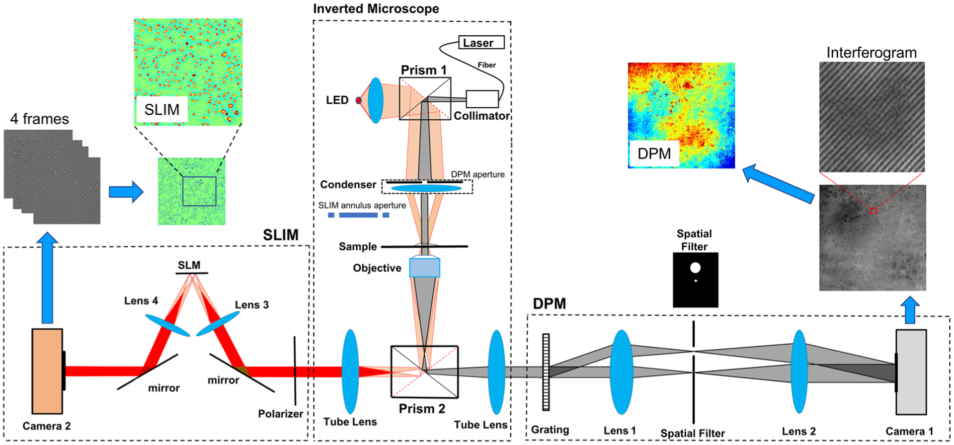
Quantitative phase imaging (QPI) has been widely applied in characterizing cells and tissues. Spatial light interference microscopy (SLIM) is a highly sensitive QPI method due to its partially coherent illumination and common path interferometry geometry. However, SLIM’s acquisition rate is limited because of the four-frame phase-shifting scheme. On the other hand, off-axis methods such as diffraction phase microscopy (DPM) allow for single-shot QPI. However, the laser-based DPM system is plagued by spatial noise due to speckles and multiple reflections. In a parallel development, deep learning was proven valuable in the field of bioimaging, especially due to its ability to translate one form of contrast into another. Here, we propose using deep learning to produce synthetic, SLIM-quality, and high-sensitivity phase maps from DPM using single-shot images as the input. We used an inverted microscope with its two ports connected to the DPM and SLIM modules such that we have access to the two types of images on the same field of view. We constructed a deep learning model based on U-net and trained on over 1000 pairs of DPM and SLIM images. The model learned to remove the speckles in laser DPM and overcame the background phase noise in both the test set and new data. The average peak signal-to-noise ratio, Pearson correlation coefficient, and structural similarity index measure were 29.97, 0.79, and 0.82 for the test dataset. Furthermore, we implemented the neural network inference into the live acquisition software, which now allows a DPM user to observe in real-time an extremely low-noise phase image. We demonstrated this principle of computational interference microscopy imaging using blood smears, as they contain both erythrocytes and leukocytes, under static and dynamic conditions.
