ACTIVE INTRACELLULAR TRANSPORT IN METASTATIC CELLS STUDIED BY SPATIAL LIGHT INTERFERENCE MICROSCOPY
Silvia Ceballos Mikhail Kandel Shamira Sridharan Hassaan Majeed Freddy Monroy Gabriel Popescu
Journal of Biomedical Optics 111209-1 (November 2015) Vol. 20(11) 2015 https://doi.org/
![]()
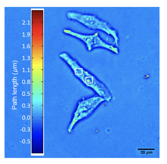
Spatiotemporal patterns of intracellular transport are very difficult to quantify and, consequently, con- tinue to be insufficiently understood. While it is well documented that mass trafficking inside living cells consists of both random and deterministic motions, quantitative data over broad spatiotemporal scales are lacking. We studied the intracellular transport in live cells using spatial light interference microscopy, a high spatiotemporal resolution quantitative phase imaging tool. The results indicate that in the cytoplasm, the intracellular transport is mainly active (directed, deterministic), while inside the nucleus it is both active and passive (diffusive, random). Furthermore, we studied the behavior of the two-dimensional mass density over 30 h in HeLa cells and focused on the active component. We determined the standard deviation of the velocity distribution at the point of cell division for each cell and compared the standard deviation velocity inside the cytoplasm and the nucleus. We found that the velocity distribution in the cytoplasm is consistently broader than in the nucleus, suggesting mechanisms for faster transport in the cytosol versus the nucleus. Future studies will focus on improving phase measurements by applying a fluorescent tag to understand how particular proteins are transported inside the cell.